 |
Gastrointestinal and Liver CenterBangkok Hospital Phuket |

|
|
Gastrointestinal and Liver CenterBangkok Hospital Phuket |
Digestive diseases or gastrointestinal diseases are the common conditions in all ages both in children and adults.
|
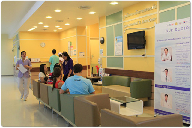
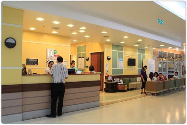
Gastrointestinal & Liver Center is located on the 1st floor, Bangkok Hospital Phuket.
The GI Center of Bangkok Hospital Phuket is a full service Gastroenterology practice exclusively specializing in the diagnosis and treatment of gastrointestinal diseases and disorders. Your comfort is a priority in our center, where you are greeted with a warm, welcoming smile. Thanks to our professional experience and know-how, we provide you with best services. Our facilities include:
- Fibroscan with CAP (Controlled Attenuation Parameter)
- Special Diagnosis without Contrast
- Ultrasonography: Imaging examination of the liver, pancreas, gall bladder, common bile duct, spleen and the kidney
- Special Diagnostic with Contrast
- Barium Swallowing: Review the swallowing and regurgitating of the esophagus
- Upper GI Examination: Examine the swallowing, esophagus, stomach and proximal part of the small intestine
- Long GI Examination: Check the swallowing, stomach, and whole small intestine
- BE (Barium Enema): Investigate the large intestine or colon
- Computerized Tomography (Spiral CT Scan) and the Magnetic Resonance Imaging (MRI) Argon Plasma Coagulation: Stop bleeding from an ulcer or abnormal vessel in the stomach and intestine through endoscopy
- Fluoroscopy: A technique for obtaining X-ray images capturing motion. The Radiologist uses a switch to control an X-Ray beam that is transmitted through the patient. The X-ray then strikes a fluorescent plate that is coupled to an “image intensifier” this in turn is connected to a television camera. The Radiologist can then watch the images “live” on a TV monitor.
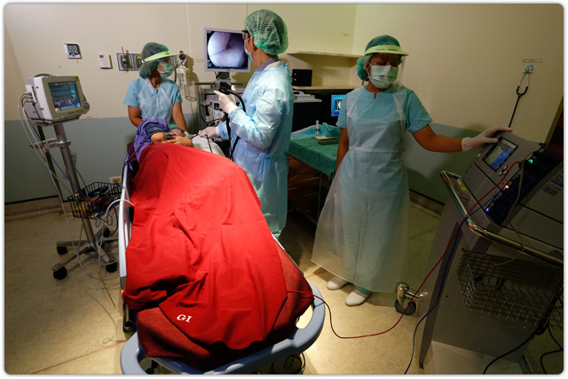
- GI Endoscopy: A visual examination of the intestinal tract using a lighted, flexible fiberoptic or video endoscope. Upper endoscopy enables the physician to look inside the esophagus, stomach, and duodenum (first part of the small intestine). The procedure might be used to discover swallowing difficulties, the causes of nausea, vomiting, reflux, bleeding, indigestion, abdominal pain, or chest pain. Upper endoscopy is known as EGD, which stands for esophagogastroduodenoscopy.
- Gastroscopy: A medical term that has two parts: gastro for “stomach” and scopy for “looking”. Gastroscopy is a diagnostic test that enables the doctor to view the stomach. The instrument used to perform this simple test is the gastroscope; a long, thin, flexible fiberoptic tube. Within the end of this remarkable device is a miniaturized color TV camera with a wide angle lens. By passing this “scope” through the stomach, your doctor can directly examine the lining of your upper digestive system. The examination is quick, painless and without incision.
- Colonoscopy: Allows the physician to investigate the entire large intestine, from the lowest part, to the rectum, all the way up through the colon to the lower end of the small intestine. This procedure is used to detect early signs of cancer in the colon and rectum. It can also be used to diagnose the causes of changes in bowel habits. Colonoscopy enables the physician to detect inflamed tissue, abnormal growths, ulcers and bleeding.
- Flexible Sigmoidoscopy: Enables the physician to assess the inside of the large intestine via the rectum and the last part of the colon, known as the sigmoid colon. Physicians may use this procedure to investigate symptoms of diarrhea, abdominal pain, or constipation. This procedure can also be adapted to detect early signs of cancer by allowing the physician to identify signs of bleeding, inflammation, abnormal growths and ulcers in the descending colon and rectum.
- Polypectomy: The removal of polyps from the stomach, small intestine and colon.
- EVS, EVL (Endoscopic Variceal Treatment): Endoscopic investigation enables the physician to treat varice in the esophagus.
- PEG (Percutaneous Endoscopic Gastrostomy): A tube enters the stomach through the abdominal wall, under the visual guidance of an endoscope.
- Diagnostic ERCP (Endoscopic Retrograde Cholangiopancreatography): Identifies a problem within the bile duct or pancreas e.g. gallstones, cancer of the bile ducts, pancreatitis.
- Therapeutic ERCP (Sphincterotomy, Stone extraction): Utilises a tube to correct a problem in the bile ducts. For example: cancer or gallstones in the bile ducts.
- GI Pathology: Extraction of cells for precise analysis.
- Interventional Radiology: Use of X-rays, ultrasound and other medical images to guide small instruments through the blood vessels or other pathways to treat disease percutaneously.
- TOCE (Transcatheter oily chemo-embolization): When the cancer cannot be removed by surgery, a radio therapist will place a small tube through the hepatic artery direct to the cancerous area, Releaseeing anticancer drugs to block the artery feeding the area. This procedure can help to reduce the cancer size and prevent it from spreading, in some cases surgery can then be used to remove it
- FNA (Fine needle aspiration) and Liver Biopsy: A liver biopsy allows the physician to examine signs of disease and damage to the liver tissue. A special needle is used to remove the tissue from the liver. A liver biopsy is carried out after tests suggest that the liver is not functioning correctly. For example, a blood test might show that your blood contains higher levels of liver enzymes or too much iron or copper, an x ray could suggest that the liver is swollen and looking at the liver tissue itself is the best way to determine whether the liver is healthy or what is causing the damage.
Click on image to view the large image.


Service Hours
Gastrointestinal & Liver Center is open daily from 8.00 am to 5.00 pm.
Contact Us
Phone: +66 7625 4425 ext. 3772, +66 7636 1000 ext. 3772


