At Radiology & Imaging Center of Bangkok Hospital Phuket, we offers a wide range of diagnostic imaging services include: Mammography, Ultrasound (2D, 3D and 4D), CTA scan, CT scan, MRI scan and CMR scan including interventional radiology services and sub-speciality expertise available under the one roof to meet all your radiological needs. Our center is staffed by dedicated and experienced radiographers and specialist consultant radiologists report all images and scans.


Mammography Information |
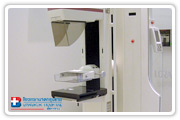 A mammogram is a special x-ray of the breast. It is a radiological procedure that can detect small cancers long before they can be felt by you or your doctor. As the x-rays pass through the breast tissue, an actual picture of the tissue inside is obtained. This image is interpreted by a radiologist who evaluates it for changes in density, the presence of calcifications or changes in the arrangement of the tissue. If you are not having any breast problems, you will be scheduled for a screening mammogram. If you are having problems, then you should be scheduled for a diagnostic mammogram.
Common uses of this procedure
Mammography is used to diagnose breast diseases in women. The use of screening mammography can assist in the detection of breast cancer even if you have no complaints or symptoms. Mammograms are recommended that women aged 40 and continuing for as long as a woman is in good health.
Women who are at an increased risk due to a genetic history of breast cancer, or who have had breast cancer, may need to get mammograms at an earlier age.
The following are suggested guidelines:
Between the ages of 35 and 40 a woman should have at least one baseline mammogram. Starting at the age of 40 a woman should have a mammogram once every year.
How does it work?
The breast is exposed to a small dose of radiation to produce an image of internal breast tissue. The image of the breast is produced as a result of some of the x-rays being absorbed (attenuation) while others pass through the breast to expose the film. The exposed film is either placed in a developing machine, producing images much like the negatives from a 35-mm camera, or images are digitally stored on computer.
The Benefits of a Mammography
Imaging of the breast can detect small tumors. When tumors are small, effective treatment and cure are more likely. The use of screening mammography increases the detection of small abnormal tissue growths confined to the milk ducts in the breast, called ductal carcinoma in situ. These early tumors cannot harm patients if they are removed at this stage and mammography is the only proven method to reliably detect these tumors.


